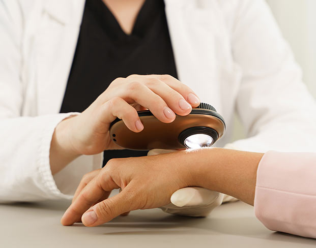
Medical

The Pure Dermatology team wants to address all of your skin concerns. We enjoy medical dermatology and will work with you to treat the common as well as more exceptional conditions you may have. Whether you have a concerning rash, new mole or a common problem like acne or warts, we are here to treat these to your satisfaction.

Please call our office today at 205.682.8022 to schedule an appointment.

Acne

Acne is the most common skin condition in the United States, affecting about 40 to 50 million Americans at any one time. Most people who are affected by acne are teenagers and young adults. However, acne can develop at any age.

Invisible
Causes
The development of acne is multifactorial, meaning that many processes lead to the formation of an acne bump.

Acne appears when a pore is clogged. This clogging process is begun by dead skin cells. Typically, dead skin cells are rise to the surface of the pore and are shed. However, when our bodies produce too much sebum (or oil), the skin cells become more “sticky”, causing them to stick together and get trapped inside the pore.

This initial clog traps all sorts of other debris inside the pore. Sometimes, p. acnes, a bacteria that naturally lives on our skin, also gets trapped inside the pore, where they have the perfect environment to multiply quickly. This encourages inflammation of the pore and promotes the development of more inflammatory acne bumps.

Excessive oil production is also a factor in the formation of acne. Oil glands are controlled by hormones in the body, which means acne often increases during times of hormonal changes like puberty and pregnancy.

Treatment
Given the multifactorial nature of acne formation, several medications or treatments may be required to treat the different aspects of formation. When you visit Pure Dermatology, our providers will develop a treatment plan that is specific to you.

Treatment types include topical treatments and medicine. Topical treatments are applied directly to your skin. Some help kill bacteria, and others reduce oil production. Medicine is sometimes necessary to treat acne, including acne cysts and nodules. Your provider may prescribe an antibiotic, birth control or other hormone related medicines (which are especially helpful for women), or Isotretinoin.

Whatever treatment plan you are given, consistency is important. Medications and topical treatments must be used consistently, or they will not be effective. Even with a great treatment plan, it usually takes several weeks for acne to start clearing up. Be patient and continue with your treatment until your next follow-up appointment. Typically, it will be 6-8 weeks before you start seeing results from your treatment.

Rosacea

Rosacea is a common skin condition, affecting approximately 10% of the population. It typically affects fair-skinned adults, but it can affect all age ranges and skin types.

Invisible
Symptoms
Rosacea often begins as a redness in the nose and cheeks. However, this redness can spread, extending to the forehead and chin, as well as beyond the face to ears, chest, and back.

There are usually some little broken blood vessels (telangiectasias) on the skin. Many people may get acne bumps with their rosacea and this is often called adult acne. Some people will get significant flushing episodes (flares of redness). Rarely, you can get an associated thickening of the skin, most commonly seen on the nose of men.

Rosacea can also affect the eyes. If you experience sensitivity to light, a gritty feeling in your eyes, frequent styes, or other new eye symptoms, please let your provider know.

Triggers
People will often be able to identify some triggers for their rosacea (things that cause flushing, make the redness worse, or cause more acne bumps).

Sunlight

Sunlight (or ultraviolet light) is one of the most common triggers of rosacea. For this reason, it is extremely important for you to wear sunscreen of at least SPF 30 every day.

Other Triggers

  • Hot foods
  • Spicy foods
  • Alcohol
  • Hot or cold temperatures

Psoriasis

Psoriasis is a chronic disease that affects approximately 2% of the population. Typically, new skin cells form over the course of few weeks. In people affected by psoriasis, skin cells form rapidly over a few days. The body does not shed this skin, so it builds up into raised pink plaques with silver-white scales.

Invisible
Types of Psoriasis

Plaque Psoriasis

Plaque psoriasis is the most common form appearing in 80-90% of patients. It appears as patches of raised, round, red skin, and can be covered in silver-white scales. These patches can occur anywhere on the skin, and may be few or may cover the entire body.

Guttate Psoriasis

Guttate psoriasis occurs in about 2% of patients and is most common in individuals under 30. It appears as small pink bumps and occurs primarily on the trunk and parts of limbs close to the trunk. Guttate psoriasis often appears a few weeks after a cold, and can take few weeks to few months to clear. If you have plaque psoriasis and develop guttate psoriasis, it means your psoriasis is worsening.

Pustular Psoriasis

Pustular psoriasis looks like white pus-filled bumps surrounded by red skin. These bumps can be all over the body, or can be on just the palms of the hands or the soles of the feet.

Inverse Psoriasis

Inverse psoriasis looks raw with red, smooth patches. These patches develop only in the folds of the skin, such as the armpits or groin.

Erythrodermic Psoriasis

Erythrodermic Psoriasis is the least common form of psoriasis and causes severe redness and shedding of the skin. Fever and flu-like symptoms are common with this type of psoriasis.

Nail Psoriasis

Nail psoriasis can appear in all psoriasis types and affects the fingernails and the toenails. It causes tiny holes in the nails, which may cause them to loosen, thicken, and eventually crumble.

Psoriatic Arthritis

Psoriasis can also cause a specific type of arthritis. The first sign is frequently swollen, stiff, and sometimes painful joints upon waking up. If you are having joint pain or arthritis symptoms, please discuss this with your provider.

Triggers

Different people have different triggers. Common triggers include:

  • Stress
  • Strep throat
  • A bad sunburn
  • Trauma to the skin
  • Smoking
  • Obesity
  • Some medications
Treatment

There are many options for treating your psoriasis. If your psoriasis is localized to a few body areas, we typically start with topical medications. For disease that persists despite topical treatments, or for more severe disease, we offer phototherapy at our office (we have both full body NBUVB and hand/foot NBUVB). Occasionally, we will need to treat psoriasis with more serious medications by mouth or with injections. These do have potential side effects which will be discussed with you. Your provider will help you to determine the best treatment plan for your psoriasis.

Skin Cancer

Skin cancer is the most common cancer, with about 1 in every 5 Americans developing it in their lifetime. If caught early, skin cancer is highly treatable.

Invisible
Skin Cancer Risks

We know that ultraviolet light plays a role in developing most skin cancers. For this reason, it is extremely important to practice sun protective measures. We recommend wearing a water-resistant sunscreen with at least SPF 30 everyday on sun exposed areas. Try to avoid being outdoors in the middle of the day. Try to stay in a covered, shady area if possible when outdoors. And sun protective clothing is a great, and now fashionable, option.

Anyone can get skin cancer, but some have a higher risk than others. Age is a key risk factor, but there are other factors. People with a higher risk for skin cancer have:

  • Light colored skin
  • Skin that burns or freckles rather than tans
  • Blonde or red hair
  • Blue or green eyes
  • More than 50 moles
  • Irregularly-shaped or darker moles

Medical history can also increase your risk of getting skin cancer. You have a great risk of skin cancer if you have:

  • History of sunburns, especially blistering sunburns
  • Used (or use) indoor tanning devices
  • Received an organ transplant
  • Had skin cancer (or have a family history of skin cancer)
  • A weakened immune system
  • Been exposed to cancer-causing compounds
  • An area of skin that has been badly burned, either in an accident or by the sun
Types of Skin Cancer

Basal Cell Carcinoma (BCC)

Basal Cell Carcinoma is the most common type of skin cancer. It appears on the skin in many shapes and sizes. It can manifest as a dome-shaped growth with visible blood vessels; as a shiny, pinkish patch; or as a sore that heals and then returns.

BCC most often develops on skin that receives a lot of sun, like the scalp, face, nose, neck, and hands. It very rarely spread to other parts of the body; however, it does continue to grow in the skin and for this reason, it is important to be treated appropriately.

With treatment, BCC is almost completely curable. Depending on the type of basal cell you have, where it is located, and the size of your cancer, your provider may recommend different treatments for you. In general, BCC is treated with an excision of the lesion, a destructive treatment called electrodessication and curettage, Mohs surgery or a topical cream.

Squamous Cell Carcinoma (SCC)

Squamous Cell Carcinoma is the second most common type of skin cancer. SCC can appear in many different shapes, including a crusted or rough bump; a red rough flat patch; a dome shaped bump that grows and bleeds; or a sore that does not heal, or heals and returns.

SCC most often develops on skin that receives a lot of sun, like the scalp, face, nose, neck, and hands. But it can also develop on areas not exposed to the sun, such as the inside of the mouth or genitals. Left untreated, SCC can spread to other parts of the body, making treatment difficult.

However, when detected early, SCC is completely treatable. For most SCCs, we recommend surgical treatment in our office or Mohs surgery, depending on the size of the cancer and the location of the SCC. Your provider will determine which method of treatment is best for you.

Melanoma

Melanoma is the deadliest form of skin cancer. It is estimated that 1 in 75 Americans will develop Melanoma in their lifetime. It may develop on normal skin or in an existing mole. Signs of melanoma include a change in the size, color, or shape of a mole, or a mole that becomes painful or begins to bleed or itch. Some melanomas form on normal skin. A new mole or growth, particularly one that does not match your other moles, may be melanoma.

When detected early, melanoma is curable. However, if melanoma grows in the skin, it can spread to your lymph nodes, lungs and other parts of your body. For this reason, early detection is extremely important.

It is important to examine your skin regularly. If you develop a new mole or you notice a mole changing in color, size, shape, etc, then you should have that lesion evaluated. If you are diagnosed with melanoma, your provider will discuss the features of your melanoma with you. Treatment and potential increased testing will depend on the features of your melanoma.

Dysplastic Nevi (Moles)

Dysplastic nevi, or atypical moles, are benign (non-cancerous) moles that can look like melanomas. Atypical moles usually have an asymmetrical shape; an irregular and/or hazy border; varied or irregular colors; and are typically larger than 6mm. Atypical moles are most commonly found on the back, chest, abdomen, and extremities, but can also occur on the buttocks, groin, breasts, or scalp.

While dysplastic nevi are not cancerous, if you have one or more atypical moles, we know that you may be at a higher risk for developing cancerous moles. For this reason, we will recommend that you have regular total body skin exams. We also recommend that you continue to monitor your moles and let us know if you are concerned about any new or changing areas.

Actinic Keratoses (AKs)

Actinic Keratoses are common skin growths that are precancerous lesions. They are often scaly, red spots that may be tender. They most commonly occur on areas that get a lot of sun exposure, like the head, including ears, lips and scalp; arms; and hands. If left alone, a small percentage of these will progress to skin cancers (squamous cell carcinomas).

We want to treat AKs before they have the opportunity to progress to skin cancer. If there are a few lesions, we often treat with liquid nitrogen (called cryotherapy). If there are numerous AKs, your provider may discuss treating the whole area to try to get rid of the AKs that are present, as well as the AKs that may be developing under the skin. This process, called field treatment, can be accomplished by several methods, usually with prescription creams or a light treatment called photodynamic therapy. Often, we will use a combination of these treatments to treat areas with lots of sun damage and AKs. Your provider will help you to develop the best plan to treat your precancerous lesions.

Signs of Skin Cancer

Early detection is key in the treatment of skin cancer, specifically melanoma. When examining your skin, follow the ABCDEs of skin cancer to detect early signs.

  • Asymmetry: A line drawn through the middle would not create matching halves
  • Border: The mole has an irregular, scalloped, or poorly defined border that fades into the surrounding skin
  • Color: The color of the mole varies or is irregular from one area to another, with shades of tan, brown, dark brown, red, blue, or black.
  • Diameter: Melanomas are generally larger than 6mm (the size of a pencil eraser) when diagnosed, but can be smaller.
  • Evolving: A mole or skin growth that looks different from the rest or changes in appearance (size, shape, or color) should be examined by your dermatologist.
How to Self-Examine Your Skin

You should examine your skin often for changes. Remember to follow the ABCDEs of skin cancer and check for anything that is changing, itching, or bleeding.

  • Examine your body (front and back) in a mirror, paying particular attention to the legs.
  • Bend your elbows and carefully examine the forearms, back of the upper arms, and palms.
  • Look at your feet, the spaces between the toes, and the soles.
  • Examine your back and scalp with a hand mirror. Remember to check your scalp by parting your hair and lifting it away from your head.
  • Check your back and buttocks with a hand mirror.
Treatment

The only way to diagnose any type of skin cancer is with a skin biopsy. Your dermatologist can perform this procedure during an office visit. To perform a skin biopsy, your dermatologist will remove the entire growth or part of it. Your dermatologist may send this to a laboratory or look at it under a microscope. The findings will be communicated in a biopsy report.

Your treatment will depend on the type and severity of your skin cancer. Your provider will discuss the available treatments and help you decide which course is right for you.

Common treatment types include excision of the lesion, electrodessication and curettage, and Mohs Surgery. Many skin cancers can be removed in our office.

However, in some cases, treatment might require a referral to another surgeon. In this case, our office will facilitate the referral for you.

Eczema

Eczema is a common skin condition that often appears as a red, scaly, itchy rash, and can occur on many different parts of the body.

Invisible
Types of Eczema

There are several different types of eczema.

Atopic Dermatitis

Atopic dermatitis is the most common type of eczema and is often described as “the itch that rashes.” Scratching can lead to redness, swelling, cracking, weeping of clear fluid, crusting, and scaling of the skin.

For most people with atopic dermatitis, it will begin during their first few years of life, with 90% of patients developing the condition before age 5. While rare, atopic dermatitis can appear at puberty or later.

While atopic dermatitis can be a lifelong condition, it often becomes less severe with age, and many children outgrow the condition all together. As the patient ages, the itchy patches will often develop on the elbow and knees, or in flexural areas such as the folds of the arms and legs. In teens and adults, patches of atopic dermatitis are typically dry, red to brownish gray, and may be scaly or have thickened skin.

Asteatotic Eczema

Asteatotic eczema is commonly seen in older adults, usually over the age of 60, though it is not unusual for adults in their 20s to develop the condition as well. It occurs when the skin becomes abnormally dry, itchy and cracked and can appear as red, dry flaky patches most commonly on the lower legs as well as the thighs, chest and arms. Asteatotic eczema is most common during the winter months, especially in areas where indoor humidity is decreased by heating.

Seborrheic Dermatitis

Seborrheic dermatitis is characterised by a red, scaly, itchy rash, most commonly found on the scalp, sides of the nose, eyebrows, ears, eyelids, and middle of the chest. It is probably related to active oil glands and also to an overgrowth of yeast that normally lives on the skin. It is a chronic rash, but can be kept under control with a good regimen.

Perioral Dermatitis

Perioral dermatitis is a rash that forms around the mouth. This rash is most common in young and middle aged women, though children and adolescents (both girls and boys) can develop this rash too. However, men rarely develop perioral dermatitis.

Periorial dermatitis appears as redness, swelling, and acne-like bumps that usually circle the mouth, leaving a think band of skin around the lips that looks normal. Many patients experience a burning sensation around the mouth. Sometimes the rash itches, and in other cases, the skin can peel and look scaly.

Treatment

For most eczemas, good skin care is of utmost importance. Further treatment will be based on the patient and the type of eczema present. Schedule an appointment with your provider to diagnose your specific type of eczema and develop a treatment plan.

Contact Dermatitis

Almost everyone gets contact dermatitis at least once in their lives. We get contact dermatitis when something that our skin touches causes a rash. Some rashes happen immediately, but most take time to appear. Rashes can be caused by an allergic reaction (allergic contact dermatitis) or can develop as a result of skin irritation.

Invisible
Allergic Contact Dermatitis
Allergic contact dermatitis is an eczema that occurs when allergy-causing substances touch your skin. If you are sensitive to them, allergens can cause an itchy rash on swollen, reddened skin that may blister, ooze, form a crust, or flake off. Poison ivy and poison oak are two common examples of contact dermatitis.

Other common allergens that can cause allergic contact dermatitis are:

  • Fragrances
  • Certain metals (like nickel in jewelry)
  • Certain topical medications
  • Some skin care products
  • Latex and other synthetic rubber products
  • Plant oils and botanical extracts, such as tea tree, sesame, cinnamon, peppermint, olive, and castor oils
Irritant Contact Dermatitis
Irritant contact dermatitis is a more common form of contact dermatitis that can look very similar to an allergic contact dermatitis. However, in this rash, you are not allergic to the substance. The rash is caused by prolonged contact with something that irritates the skin.

Common types of irritant contact dermatitis are:

  • Diaper rash
  • Acid burn
  • Dry, cracked hands due to frequent hand washing
  • Irritated skin around the mouth due to lip licking
  • Pepper spray

Several professions are especially susceptible to irritant contact dermatitis, including beauticians, nurses, bartenders, and others who spend lots of time with wet hands.

If your rash does not clear up within a few weeks, please consult your provider. When contact dermatitis develops, treatment is important. It can prevent the contact dermatitis from worsening and help your skin heal.

Patch Testing
Patch testing (a type of allergy testing usually performed by a dermatologist) can be helpful to identify certain allergens that may be responsible for the rash.

Common Skin Growths

There are several common types of skin growths. These growths are benign, meaning they are non-cancerous. However, if you have any concerns about your growth, do not hesitate to ask your provider about it.

Warts

Warts are benign skin growths that are caused when the human papillomavirus (HPV) infects skin cells. You are more likely to get one of these viruses if you cut or damage your skin in some way. Warts are contagious and can be spread by contact with the wart or something that touched the wart.

Invisible
Types of Warts
Warts can look a lot of different ways, and this often depends on the type of wart.

  • Common warts most often grow on the fingers, around the nails, and on the backs of hands. They are more common where skin was broken, such as from biting fingernails or picking at hangnails and most often feel like rough bumps.
  • Plantar warts grow most often on the soles of the feet. They are often flat or grow inward and sometimes grow in clusters. A plantar wart can hurt, and it may feel like you have a pebble in your shoe.
  • Flat warts can occur anywhere. Children usually get them on the face, while men get them most often in the beard area, and women get them on their legs. Flat warts are smaller and smoother than other warts and tend to grow in large numbers (20 to 100 warts at a time).
Treatment
In most people (especially kids), the immune system will eventually clear the virus and the warts will resolve. However, it can sometimes take several months to years for the immune system to clear the warts. There is a smaller group of people that tend to keep persistent warts, meaning that the warts are never cleared by the immune system.

Warts are often very difficult to treat; however, there are many different options available to try to clear them. No treatment works for everyone and it usually takes multiple visits to resolve the warts completely. Your provider will determine the best treatment option(s) for you.

Moles

Moles are extremely common. In fact, almost every adult has a least a few moles. People with light skin tend to have more moles, sometimes 40 or more.

Most moles appear during the first 20 years of life, though some may not form until later. Sun exposure increases, and sometimes darkens, moles. During the teen years and pregnancy, moles may get larger and darker, and new ones may appear.

Invisible
Types of Moles
Moles can appear anywhere on the skin, and come in all shapes and sizes.

  • Common/Normal Moles: Common moles are usually brown in color, but they can also be skin color. They may be flat and tan like a freckle, or they can be raised from the skin and very noticeable. They can even contain dark hairs.
  • Congenital Moles: A congenital mole is a mole that a person has from birth. About 1 in every 100 people will be born with a congenital mole. These moles can range in size from small to giant. Having a congenital mole increases a person’s risk of developing melanoma.
  • Atypical Moles: Dysplastic nevi, or atypical moles, are benign (non-cancerous) moles that can look like melanomas. Atypical moles usually have an asymmetrical shape; an irregular and/or hazy border; varied or irregular colors; and are typically larger than 6mm. Atypical moles are most commonly found on the back, chest, abdomen, and extremities, but can also occur on the buttocks, groin, breasts, or scalp. People with atypical moles may be at a higher risk for skin cancer and should be sure to self-examine their skin regularly.
Signs of Skin Cancer
People with atypical moles are at a higher risk for developing skin cancer. Early detection is key in the treatment of skin cancer, specifically melanoma. When examining your skin, follow the ABCDEs of skin cancer to detect early signs.

  • Asymmetry: A line drawn through the middle would not create matching halves
  • Border: The mole has an irregular, scalloped, or poorly defined border that fades into the surrounding skin
  • Color: The color of the mole varies or is irregular from one area to another, with shades of tan, brown, dark brown, red, blue, or black.
  • Diameter: Melanomas are generally larger than 6mm (the size of a pencil eraser) when diagnosed, but can be smaller.
  • Evolving: A mole or skin growth that looks different from the rest or changes in appearance (size, shape, or color) should be examined by your dermatologist.

Seborrheic Keratoses (SKs)

Seborrheic keratoses (SKs) are common, benign skin growths that can occur anywhere on the skin. They most often form on the chest, back, scalp, face, and neck. They do not form on the palms of the hands or soles of the feet. While anyone can develop an SK, they are typically associated with old age. These growths generally first appear in middle age or later. Children rarely develop SKs.

Invisible
Appearance
Most SKs begin as a small, rough bump, though they can also be smooth and flat, and sometimes are mistaken for other common growths like warts or moles. They are typically brown, and range in color from tan to black. The most distinguishing trait of a seborrheic keratosis is its dull, waxy, pasted-on-the-skin appearance. An SK will often look like a dab of candle wax on the skin.

Treatment
As seborrheic keratoses are benign, treatment is usually not necessary. However, your provider might recommend removing an SK that grows quickly, turns black, itches, or bleeds, as these are signs of potential skin cancer.

Though SKs are benign, some people ask to have them removed, especially if they are large, easily irritated by clothing or jewelry, or are considered unsightly.

Molluscum

Molluscum contagiosum is a common skin disease that is caused by a virus. It appears as small, pink or flesh-colored bumps on the skin. The bumps have a dimple in the center that may or may not be filled with a thick, white substance. Though molluscum bumps are painless, some of the bumps may be itchy. Molluscum lesions will turn red as the immune system fights the virus. It is important to note that the virus can appear on other areas of the body after a person scratches or picks the bumps. Scratching or picking can spread the virus.

Molluscum is seen most often in children, but people who live in a tropical climate are also likely to contract the virus, as it thrives in warm, humid places.

Invisible
Causes
Molluscum is spread by a virus. There are 2 ways to contract the mollusucm contagiosum virus.

  • Touching something infected with the virus: The virus can be spread by sharing infected towels or clothing. Wrestlers and gymnasts can also contract the virus by touching an infected mat.
  • Direct skin-to-skin contact: Children often contract molluscum from touching other children with the virus. Teens and adults often contract the virus through sexual contact.
Treatment
Molluscum is a viral infection that the immune system will eventually recognize. However, it may take years for the immune system to clear these lesions.

When multiple lesions are present, your provider may recommend treatment. There are various treatment options available for molluscum. When multiple lesions are present, several treatment sessions are often required to completely clear all of the bumps. It is important to realize that mild scarring may occur from these lesions with or without treatment.

Urticaria (Hives)

Urticaria, or hives, are raised, itchy red welts that can appear on any part of the skin. Hives vary in size and can be as small as a pen tip or as large as a dinner plate. They may even connect to form larger welts.

When large welts occur deeper under the skin, the medical term is angioedema. Angioedema can occur with hives, and often causes the eyelids and lips to swell. If this occurs, you should seek emergency care right away, especially if you are having difficulty breathing. The vast majority of hives are only in the skin and not associated with these concerns.

A hive often lasts less than 24 hours, but new hives may appear as old ones disappear, so hives may last a few days or longer. All hives are considered acute at first, meaning they last less than 6 weeks. If a bout of hives lasts longer than 6 weeks, they are considered to be chronic hives.

Invisible
Causes
Common causes of acute urticaria are an allergy, infection, drug, or food, but many cases have an unknown cause. Some cases have a pretty obvious trigger (for example a certain food or a new medication). Hives typically occur within hours of consuming a culprit food or within a few days of starting a new medication. In kids especially, a virus/ infection may cause hives to occur. Sometimes the illness was so mild that you didn’t know you were sick. Most often, we never identify or find the cause of acute hives, but they eventually just go away.

Chronic urticaria (hives lasting longer than 6 weeks) can be associated with some medical conditions; however the vast majority of chronic hives are idiopathic. This means that even with extensive testing we are not able to identify a cause for the hives. Often we are left managing the hives until they hopefully resolve. Chronic hives may last for years.

Treatment
Antihistamines are the main treatment for most patients with hives. These medications can reduce itching, flatten the raised welts, and potentially shorten the duration of the rash. There are several types of antihistamines that your provider might recommend. Long acting antihistamines like Allegra, Claritin, and Zyrtec, are less likely to cause drowsiness. Shorter acting antihistamines like Benadryl provide faster relief, but wear off quickly and can cause drowsiness. Your provider might recommend treatment with several antihistamines at the same time. In general, antihistamines should be taken on a DAILY basis as recommended by your provider. They should not be taken only when you are breaking out or symptomatic.

Certain things may tend to make hives worse – heat and stress for example. Aspirin and other NSAIDs can make hives worse and should be avoided if possible.

If hives still occur even with numerous antihistamines, we may discuss blood work or other options for treatment. Xolair is an injection that can be given for chronic idiopathic urticaria. Idiopathic urticaria is the most common type of chronic urticaria, and it is when the cause of the rash is unknown. This shot is given by the allergist and used in people who have hives not controlled by antihistamines or other medications.

Dry Skin

Dry skin is a common condition that can can occur at all ages. It can cause rough, scaly, or flaking skin; itching; and even cracks in the skin, which may bleed if severe. Sometimes people need a dermatologist’s help to get relief from dry skin. Extremely dry skin can be a warning sign of a skin problem called dermatitis. Dermatitis means inflammation of the skin. It can cause an itchy rash or patches of dry irritated skin. The earlier dermatitis is diagnosed and treated the better. Without treatment, dermatitis often gets worse.

Invisible
Causes
Anyone can get dry skin. Skin becomes dry when it loses too much water or oil. Some causes of dry skin are:

  • Age: Glands in our skin deposit oil onto the skin surface that helps to keep the skin hydrated. As we age, the number of oil glands in our skin decreases and thus makes the skin more susceptible to becoming dry.
  • Climate and/or Weather: Places with a very low humidity, such as dry, desert climates, or heated indoors during the winter can contribute to dry skin.
  • Skin Disease: People who had atopic dermatitis (also called eczema) as a child tend to have dry skin as adults. Psoriasis also causes very dry skin.
  • Excessive Hand Washing: Excessive bathing, hand washing, or scrubbing can strip the oils from your skin, which leads to dry skin.
  • Job: Nurses, hair stylists, and people in other occupations often immerse their skin in water throughout the day. This can cause the skin to become dry, raw, and cracked.
Caring for Your Dry Skin
The best way to care for your dry skin is to establish a good skin care regimen. Good skin care may require you to develop a new routine, but it will be well worth it in the long run.

The following are some important measures to help with preventing and treating dry skin and eczema rashes:

  • Take only warm baths and showers. Do not take a hot shower.
  • Use a mild, fragrance free soap or a soapless cleanser such as Cetaphil or Cerave.
  • Soap should be applied with your hands only to dirty areas (underarms, feet, groin). In general, the shower/ bath water is enough to wash the rest of your body.
  • After bathing, PAT dry with a towel. Do not rub the skin roughly with a towel.
  • Apply a good moisturizing cream (not lotion) twice daily, once immediately after bathing and again sometime during the day.
  • Your provider may recommend an over the counter lactic acid lotion.
  • Switch to a fragrance free detergent. You may also try double rinsing your clothes.
  • Avoid fabric softeners and dryer sheets.
  • Avoid all fragranced products such as body lotions and perfumes.
  • Try not to scratch. Scratching often makes the itching worse and can lead to a cycle of itching and scratching.
  • Use a humidifier at night, especially during the winter, to help replenish moisture in the air.
  • Do not use rubbing alcohol, Lanacaine, Neosporin, Caladryl, Calamine lotion, Clorox, or Lysol on your skin. These products can be drying and/or irritating and often make your condition worse.

Hair Loss

In a typical hair growth pattern, about 10% of your hair will be in a resting (telogen) state. At the end of this phase, which last two to three months, the hair falls out. When you shed hair, a a new hair grows from the same hair follicle. It is normal to lose 50 to 100 hairs per day. However, if you have bald patches or notice your hair thinning, you may be experiencing hair loss.

Invisible
Types of Hair Loss

Hereditary Hair Loss

The most common type of hair loss is hereditary hair loss, which may also be known as male-pattern baldness, female-pattern baldness, or androgenetic alopecia. About 80 million men and women in the United States experience this type of hair loss. Men may see a receding hairline and bald patches, especially on the top of their head. Women do not go fully bald, but they may see thinning hair and a wider part when they style their hair.

There is no cure for hereditary hair loss, but there are medical treatments that may help some people, like Minoxidil and Finasteride.

Telogen Effluvium

Sometimes, illness, stress, or other events can cause too many hairs to enter the resting state of the hair growth cycle. This leads to excessive shedding and thinning of hair. Typically, bald patches do not appear.

There is no treatment for telogen effluvium, but in many cases, it tends to go away after one of the potential causes is eliminated. Some causes of this type of hair loss are:

  • Childbirth or menopause
  • High fever, serious infection, or a bad bout of the flu
  • Major surgery or chronic illness
  • Thyroid disease
  • A diet lacking in iron or protein
  • Birth control pills or other medicines
  • Cancer treatments

Alopecia Areata

Alopecia areata is an autoimmune disorder that causes smooth, round patches of hair loss. It may lead to total loss of scalp hair, and can sometimes affect body hair as well. This type of hair loss can affect people of any age, including children.

In most cases, the hair will begin to regrow over time. Your provider might recommend treatments like cortisone injections or topical medications to help you regrow your hair more quickly.

Cicatricial (Scarring) Alopecia

This rare disease develops in otherwise healthy people. The disease destroys a person’s hair follicles. Scar tissue forms where the follicles once were, so the hair cannot re-grow. Treatment tries to stop the inflammation, which destroys the hair follicles.

Tinea Capitis

A fungus that can cause scaly patches on the scalp along with broken hair, redness, swelling, and even oozing on the scalp. This fungus is very contagious and is most common in children.

Treatment

The treatment your dermatologist recommends will depend on the reason for your hair loss and the health of your remaining hair.

Non-Prescription Treatment

  • Minoxidil: This medicine is applied to the scalp. It can stop hairs from getting thinner and stimulate hair growth on the top of the scalp. It is the only hair regrowth product approved for men and women with adrogenetic alopecia. A dermatologist may combine minoxidil with another treatment.
  • Laser devices: Brushes, combs, and other handheld devices that emit laser light might stimulate hair growth. The long-term effectiveness and safety for these devices are not known.

Prescription Medicine

  • Finasteride: It comes in pill form and helps slow hair loss in about 88% men. It helps stimulate hair re-growth in about 66% men. Finasteride works by stopping the body from making a male hormone, dihydrotestosterone (DHT).
  • Corticosteroid: If your hair loss is caused by inflammation in your body, a dermatologist may inject a corticosteroid into your scalp. This can help stop the inflammation that happens when a person has alopecia areata and other types of hair loss.

Hair Restoration Procedures

  • Hair transplantation: Skin on the scalp that has good hair growth is removed and transplanted to areas of the scalp that need hair.
  • Scalp reduction: Bald scalp is surgically removed and hair-bearing scalp is brought closer together to reduce balding. Scalp reduction surgery can be performed alone or in conjunction with a hair transplant.
  • Scalp expansion: Devices are inserted under the scalp for about 3 to 4 weeks to stretch the skin. This procedure may be performed before a scalp reduction to make the scalp more lax. It also can be performed solely to stretch hair-bearing areas, which reduces balding.
  • Scalp flaps: A hair-bearing segment of scalp is surgically moved and placed where hair is needed.

Granuloma Annulare

Granuloma annulare is a raised, bumpy, or ring shaped lesion. It is most common in children and young adults, though it can affect people of all ages. Women are more likely than men to develop granuloma annulare.

Granuloma annulare can occur singly or in groups on the skin. Lesions may be pearly white, skin-colored, red, or purple. Granuloma annulare typically begins as a round, firm smooth bump and then becomes a circular ring with a clear center. Lesions can develop on any part of the body, but they typically form on the hands and feet, elbows, and knees. They normally don’t peel or itch, and often can go unnoticed.

Invisible
Treatment
Since most people with granuloma annulare don’t present symptoms, treatment is not always necessary. Typically, the lesions will disappear on their own without leaving scars. Granuloma annulare normally takes a few months to clear up, but occasionally, clearing can take a few years.

If lesions are widespread or cosmetically undesirable, there are several treatments available, such as steroid creams or injections, ultraviolet light therapy, or oral medications.

Tinea Versicolor

Tinea versicolor is a common skin condition that is caused by an overgrowth of the yeast that lives on everyone’s skin. It is considered to be a fungal infection because yeast is a type of fungus. Unlike other fungal infections, tinea versicolor is not contagious.

The first sign of tinea versicolor is usually small, light-colored spots on the skin. These spots may be tan, pink, or very faint. As the yeast continues to grow, spots might combine and form patches of skin that are either lighter or darker than normal.

Anyone can have tinea versicolor, though people with oily skin, such as teens and young adults, get tinea versicolor more frequently.

Invisible
Treatment

Before recommending treatment, your dermatologist will consider several factors, including where the tinea versicolor appears on your body, how much of your skin is affected, and the weather where you live. Although the yeast is easy to control and does not cause scarring, it can take weeks or months for your skin to return to its normal color.

Topical

Your provider may recommend a prescription-strength or over-the-counter anti-fungal shampoo, soap, cream, or lotion to keep the yeast under control. The active ingredient in these products is usually selenium sulfide, ketoconazole, or pyrithione zinc.

Medicated Cleansers

Tinea versicolor is most common when it is warm and humid outdoors. During these times, a medicated cleanser with selenium sulfide, ketoconazole, or pyrithione zinc, used once or twice a month, may help to keep yeast growth under control.

Oral Medications

If your tinea versicolor covers a large area of your body or returns frequently, your dermatologist may prescribe anti-fungal pills.

Nail Fungus

Nail fungus is a common skin condition and can affect both fingernails and toenails. It is an easy infection to contract and is easily spread. If you have a fungal infection on one of your nails, it can spread to one or more of your nails.

Invisible
Symptoms

Nail fungal infections have multiple symptoms. You may have one or more of the following changes to your nails:

  • Part of the nail turns white, yellow, brown, or another color. At first, it may just be the tip of the nail that is discolored, but left untreated, will spread to the rest of the nail.
  • Debris builds up under the nail.
  • A nail begins to lift up so that it’s no longer firmly attached to the nail bed.
  • The nail turns white, and feels soft, dry, and powdery. It may even thin to the point where you can scrape off the nail.
  • Nails thicken and turn yellow or brown. This often affects all of the fingernails.
  • A nail splits or crumbles.
Causes

Nail fungus is also spread through contact of infected surfaces, especially in a warm, moist place like a pool deck or locker room. If someone with a fungal infection walked barefoot through the area, all you have to is is walk barefoot in the same area to contract the infection. Having wet nails for a prolonged period of time also makes you susceptible to a nail fungal infection. Some people develop a fungal infection from wearing the same pair of sweaty shoes or boots every day.

Nail fungus can also be spread from skin-to-skin contact from someone who has another fungal infection like athlete’s foot or ringworm on their hands.

Fungal infections can also be spread by sharing an infected pair of nail clippers or towel.

The fungi usually infect a nail by getting into a:

  • Small cut in the skin surrounding your nail
  • Crack in your nail
  • Separation between the nail and finger (or toe)

The area under a nail gives fungi a warm, moist place to grow. The infection can then spread to other nails and even your skin.

Treatment

Some treatments may begin with your provider trimming part of the nail to confirm that you have nail fungus and not some other type of nail abnormality. They may also remove some of the debris underneath the nails.

Your dermatologist may recommend one of several of the following treatments to cure your nail fungus.

Topical Medication

If your infection is mild, your dermatologist may recommend a treatment that is applied directly to the nail. This treatment helps keep new fungus out while the nail grows. Fingernails typically grow out in 4 to 6 months. Toenails take longer, usually takes 12 to 18 months.

Oral Medication

If you have a more aggressive infection, your provider might prescribe an oral antifungal pill, which has a higher cure rate and work more quickly than topical medications. An antifungal pill can cure a fingernail infection in 2 months and a toenail infection in 3 months.

Nail Removal

If you fungal infection is severe enough that the topical and/or oral medications don’t work, your provider may recommend removing the nail entirely to get rid of the infection. Your dermatologist will refer you to another provider who can perform this procedure. After the treatment, your nail will grow back.

Vitiligo

Vitiligo is a condition that causes the skin to lose its pigmentation or color. Some people only develop a few spots that may lighten or turn completely white. Others might experience a widespread loss of color.

Vitiligo usually affects the skin, but it can develop anywhere we have pigment. Patches of hair can turn white. Some people lose color inside their mouths. Even an eye can lose some of its color. While vitiligo can affect all parts of the skin, it typically begins on the hands, forearms, feet, and face. Vitiligo can affect all races and genders, but it more noticeable in people with darker skin.

Invisible
Types of Vitiligo

Generalized Vitiligo

Generalized vitiligo is the most common type. Often generalized vitiligo starts with a rapid loss of pigmentation and typically appears first on the hands, fingertips, wrists, around the eyes or mouth, or on the feet. Cycles of pigment loss, followed by times with no pigment change may continue through the person’s life. Generalized vitiligo can spread to the face, lips, hands, arms, or genitals.

Generalized vitiligo is often referred to as bilateral vitiligo because it causes loss of skin pigmentation on both sides of the body.

Segmental Vitiligo

Segmental vitiligo is called such because pigmentation loss tends to be confined to one segment of the body. A segment of hair on the head, an eyelash, or an eyebrow may turn white, and is almost always on just one side of the body. This type of vitiligo begins at an early age and only progresses for a year or two.

Trichrome Vitiligo

Trichrome vitiligo is characterized by the development of three shades of color – brown, tan, and white – on the skin.

Treatment

Light Therapy

Light therapy uses lights to repigment the skin. During this treatment, a patient may sit in a light box (which is used for widespread vitiligo) or receive excimer laser treatments (which are used to treat small areas). Light therapy works best on the face and is effective for many patients, with about 70% seeing results with the excimer laser. However, results can disappear when treatment is stopped.

Creams and Ointments

Your dermatologist may prescribe a topical medicine like corticosteroids, tacrolimus, and calcipotriol to apply directly to the skin. This treatment tends to be more effective in people with darkly pigmented skin and often is most effective on the face.

Depigmentation

For patients with widespread vitiligo, depigmentation is a treatment that might be recommended. This involves removing the remaining pigment from the skin to achieve a single skin color. This treatment is considered permanent.

Camouflage

Some patients with vitiligo prefer to camouflage their condition. For people with fair-skin, this can be done fairly successfully by avoiding tanning, which can make areas of vitiligo barely noticeable. Others camouflage their vitiligo with things like makeup, self-tanner, or dye.